Home » Without Label » Left Hip Muscles Anatomy : Tendinitis And Bursitis Treatment Cincinnati Tendinitis Dayton Oh - Learn their anatomy efficiently and easily using kenhub's muscle anatomy and reference charts!
Left Hip Muscles Anatomy : Tendinitis And Bursitis Treatment Cincinnati Tendinitis Dayton Oh - Learn their anatomy efficiently and easily using kenhub's muscle anatomy and reference charts!
Left Hip Muscles Anatomy : Tendinitis And Bursitis Treatment Cincinnati Tendinitis Dayton Oh - Learn their anatomy efficiently and easily using kenhub's muscle anatomy and reference charts!. The muscles are broken down into three layers, and are primarily used to assist with the breathing process. The semitendinosus and long head of the biceps femoris are shown on the right side; Functionally, the hip joint enjoys a very high range of motion. Left hip muscles anatomy : These ligaments reinforce and stabilize the hip joint(6).
These are gracilis, pectineus, adductor longus, adductor brevis, adductor magnus, and adductor minimus muscles. Anatomy of the hip muscles hip muscle anatomy is a complex topic. Anatomy it band pelvis muscle pelvis with muscles hip muscles muscles of pelvis tensor fascia latae psoas major anatomy pelvis tensor fascia lata pelvis muscles. These muscles include the gluteus maximus muscle (the largest muscle in the body) and the hamstrings group, which consists of the biceps femoris, semimembranosus, and semitendinosus muscles. The sartorius muscle is a distinctively long and thin muscle that crosses the thigh diagonally.
Hip Flexors from i.pinimg.com Rectus femoris muscle, one of. The abductor muscles of the hip were studied by using the variations in individual and composite muscular anatomy were recorded. Blood vessels and nerves of the hip Ligaments are soft tissue structures that connect bones to bones.a joint capsule is a watertight sac that surrounds a joint.in the hip, the joint capsule is formed by a group of three strong ligaments that connect the femoral head to the acetabulum. Learn their anatomy efficiently and easily using kenhub's muscle anatomy and reference charts! When the hip muscles are left more or less intact, they are able to support the new. The movements that can be carried out at the hip joint are listed below, along with the principle muscles responsible for each action: The deeper semimembranosus and short head of the biceps femoris are shown on the left side.
Semimembranosus, semitendinosus and biceps femoris (the hamstrings)
Blood vessels and nerves of the hip There are also diseases and disorders that can cause the pain to. Lateral rotation is needed for crossing the legs. The hamstring group is composed of the semitendinosus and semimembranosus medially and the biceps femoris laterally. The quadriceps group of four muscles. The six hip adductor muscles are all located in the adductor or medial compartment of the thigh and all mainly adduct the thigh at the hip joint. The movements that can be carried out at the hip joint are listed below, along with the principle muscles responsible for each action: 1 hip anatomy, function and common problems. Learn their anatomy efficiently and easily using kenhub's muscle anatomy and reference charts! The muscles in this region move the lower limb in the hip joint and are important muscles for. The strong muscles of the hip region also help to hold the hip joint together and prevent dislocation. This is because there are so many different muscles that give our hip joints a full range of motion. Adductor muscles on the inside of your thigh.
The muscles in this region move the lower limb in the hip joint and are important muscles for. Semimembranosus, semitendinosus and biceps femoris (the hamstrings) The iliofemoral, pubofemoral, and ischiofemoral ligaments represent the thickenings of the joint capsule. Blood vessels and nerves of the hip These ligaments reinforce and stabilize the hip joint(6).
Muscle Anatomy Of The Hips Buttocks Everything You Need To Know Dr Nabil Ebraheim Youtube from i.ytimg.com The femur may also rotate around its axis about 90 degrees at the hip. Patient education | concord orthopaedics. Left hip muscles anatomy : When the hip muscles are left more or less intact, they are able to support the new. Ligaments, tendons, and muscles play an important role in the function of the hip. Your body has two iliopsoas muscles: Gently lower your left leg on the floor. The strong muscles of the hip region also help to hold the hip joint together and prevent dislocation.
The quadriceps muscles are four powerful muscles at the front of the thigh involved in movement.
The six hip adductor muscles are all located in the adductor or medial compartment of the thigh and all mainly adduct the thigh at the hip joint. The sartorius muscle is a distinctively long and thin muscle that crosses the thigh diagonally. The abductor muscles of the hip were studied by using the variations in individual and composite muscular anatomy were recorded. Gently lower your left leg on the floor. This is because there are so many different muscles that give our hip joints a full range of motion. The femur may also rotate around its axis about 90 degrees at the hip. The hip's unique anatomy enables it to be both extremely strong and amazingly flexible, so it can bear weight and allow for a wide range of movement. Also, the posterior hip muscles produce a movements within the hip joint, such are abduction, adduction, outward rotation and retroversion. The pectineus muscle is a flat, quadrangular muscle that lies at the top of your inner thigh, often referred to as your groin muscle. Blood vessels and nerves of the hip Together, they form the part of the pelvis called the pelvic girdle. 1 hip anatomy, function and common problems. The posterior muscle group is made up of the muscles that extend (straighten) the thigh at the hip.
Anatomy of the hip muscles hip muscle anatomy is a complex topic. There are two hip bones, one on the left side of the body and the other on the right. The hamstring group is composed of the semitendinosus and semimembranosus medially and the biceps femoris laterally. The strong muscles of the hip region also help to hold the hip joint together and prevent dislocation. Functionally, the hip joint enjoys a very high range of motion.
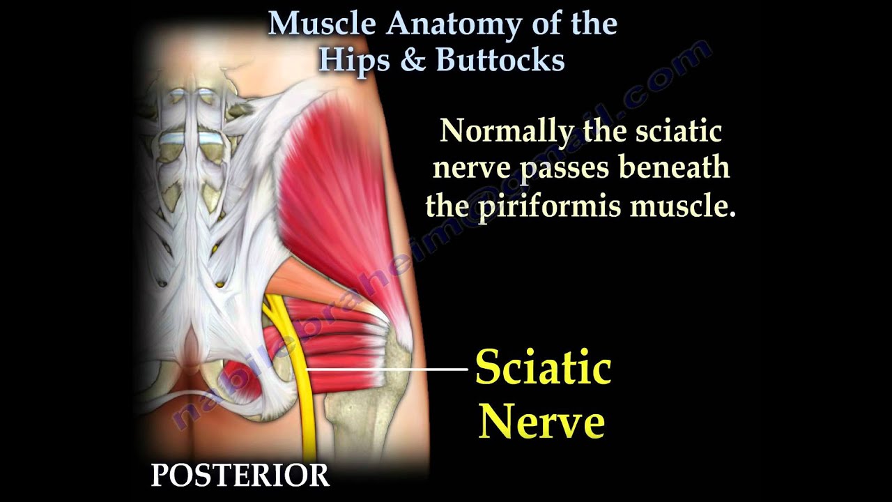
Superficial Left And Deep Right Muscles Around The Hip Download Scientific Diagram from www.researchgate.net The posterior hip musculature comprises a group of muscles extending from the pelvic bone to the femur.these muscles are important for the stabilization of the pelvis during constant mechanical stress that it suffers. In utero fetal hips lie typically in flexion, abduction and external rotation, with the left hip usually muscular anatomy. It's primarily responsible for hip flexion, but it also rotates your thigh and adducts, which means it pulls your legs together when the muscles contract. He is an attending emergency medicine phys. Functionally, the hip joint enjoys a very high range of motion. The hip muscles are composed of multiple flexors, extensors, adductors, abductors, and rotators that work together. See anatomy hip muscles stock video clips. Anatomy it band pelvis muscle pelvis with muscles hip muscles muscles of pelvis tensor fascia latae psoas major anatomy pelvis tensor fascia lata pelvis muscles.
The muscles are broken down into three layers, and are primarily used to assist with the breathing process.
Iliopsoas muscle, a hip flexor muscle that attaches to the upper thigh bone. These ligaments reinforce and stabilize the hip joint(6). Semimembranosus, semitendinosus and biceps femoris (the hamstrings) For detailed anatomy of pelvic bones, read anatomy of hip bone. The muscles in this region move the lower limb in the hip joint and are important muscles for. Patient education | concord orthopaedics. Use the mouse scroll wheel to move the images up and down alternatively use the tiny arrows (>>) on both side of the image to move the images.>>) on both side of the image to move the images. Injury to the iliopsoas may cause hip pain and limited mobility. The six hip adductor muscles are all located in the adductor or medial compartment of the thigh and all mainly adduct the thigh at the hip joint. In utero fetal hips lie typically in flexion, abduction and external rotation, with the left hip usually muscular anatomy. The view on the left has the rectus femoris cut away to show the vastus intermedius which is below it. To put it plainly, sometimes hip pain comes from the hip, but a lot of times hip pain comes from the back. Together, they form the part of the pelvis called the pelvic girdle.